
ORTHOPEDICS + SPORTS MEDICINE

Orthopedic Knee Specialists

Expert care for knee pain, instability, and arthritis, from first evaluation through recovery.

The knee is one of the body's most important weight-bearing joints, providing the stability, strength, and mobility needed for everyday life. Because it absorbs significant forces with every step, the knee is particularly vulnerable to injury, wear and tear, and age-related degeneration that can affect comfort and movement.

At Sforzo | Dillingham | Stewart Orthopedics + Sports Medicine, our team provides expert orthopedic care for patients experiencing knee pain, stiffness, instability, and loss of function. Through personalized treatment plans and advanced orthopedic solutions, we help patients restore mobility, regain confidence, and return to the activities they enjoy most.

Specialized Knee Care

Sports Medicine Knee Care

Personalized Care Plans

Understanding Knee Anatomy

The knee is a complex joint made up of bones, tendons, ligaments, and muscles, all working together to ensure proper function and stability during movement. It primarily consists of the femur (thighbone) and tibia (shinbone), with the patella (kneecap) protecting the joint.

Articular cartilage covers the bone surfaces to reduce friction, while the menisci act as shock absorbers. Ligaments, including the medial and lateral collateral ligaments and the anterior and posterior cruciate ligaments, provide stability. Major muscle groups, like the quadriceps and hamstrings, facilitate knee movement through their associated tendons.
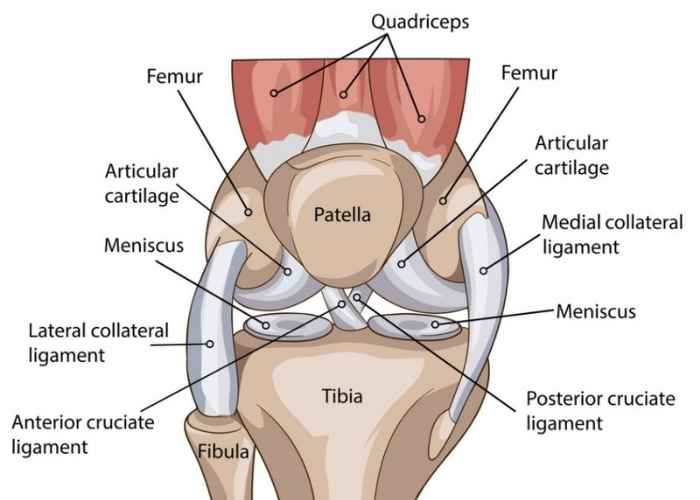
Full knee anatomy diagram

Knee Arthritis: Causes, Types, and Symptoms

Knee arthritis is a condition where the cartilage in the knee joint breaks down, leading to pain, stiffness, and limited mobility. It can significantly impact daily activities such as walking, climbing stairs, or standing for long periods.

Causes of Knee Arthritis

The causes of knee arthritis can vary, but common factors include:

  • Osteoarthritis (OA): The most common form of knee arthritis, caused by the gradual wear and tear of cartilage over time, often associated with aging or joint overuse.
  • Rheumatoid Arthritis (RA): An autoimmune condition that causes chronic inflammation in the knee joint, leading to pain, swelling, and possible joint damage.
  • Post-Traumatic Arthritis: Develops after an injury, such as a knee fracture or ligament tear, that disrupts the integrity of the knee joint and leads to arthritis over time.
  • Infectious Arthritis: Caused by an infection in the knee joint, leading to inflammation, swelling, and pain.

Types of Knee Arthritis

  • Osteoarthritis (OA): The most common form of knee arthritis, often affecting older adults. It involves the gradual loss of cartilage, resulting in bone-on-bone contact and pain.
  • Rheumatoid Arthritis (RA): An autoimmune disorder that causes inflammation in the synovium of the knee joint, often leading to pain, swelling, and deformity.
  • Post-Traumatic Arthritis: A form of arthritis that occurs after a significant knee injury or trauma, causing damage to the cartilage and accelerating the degeneration of the joint.
  • Infectious Arthritis: Rare but serious, this type of arthritis occurs when bacteria or viruses infect the knee joint, leading to inflammation and severe pain.

Symptoms of Knee Arthritis

  • Pain: Pain in the knee, especially with movement, weight-bearing activities, or prolonged sitting.
  • Stiffness: Limited range of motion, particularly after rest or in the morning.
  • Swelling: Inflammation and puffiness around the knee joint, often causing discomfort.
  • Instability: A feeling of weakness or giving way in the knee, especially with standing or walking.
  • Crepitus: A sensation of grinding, popping, or crackling in the knee joint during movement.

Knee arthritis can significantly affect quality of life, but treatment options such as physical therapy, medications, injections, or in severe cases, knee replacement surgery, can help reduce pain and improve function.

Treatments

Bursitis: Causes, Types, and Symptoms

Bursitis is the inflammation of a bursa, a small, fluid-filled sac that acts as a cushion between bones and soft tissues like muscles and tendons. When a bursa becomes irritated or inflamed, it can cause pain and swelling, making movement difficult.

Causes of Bursitis

Bursitis often results from repetitive motion, prolonged pressure, or injury to the affected area. Common causes include:

  • Repetitive Movements: Activities that involve repetitive motion, such as sports or manual labor, can lead to bursitis.
  • Trauma or Injury: A direct blow or injury to a joint can cause inflammation in the bursa.
  • Overuse: Excessive use of certain joints, especially in positions that put pressure on the bursa, can lead to irritation.
  • Infection: In rare cases, a bacterial infection can cause septic bursitis, leading to more severe symptoms.
  • Medical Conditions: Conditions like rheumatoid arthritis, gout, or diabetes can increase the risk of developing bursitis.

Common Types of Bursitis

  • Shoulder Bursitis: Inflammation of the bursa in the shoulder, often caused by repetitive overhead movements.
  • Elbow Bursitis: Also known as “student’s elbow,” it occurs when the bursa at the tip of the elbow becomes inflamed, typically from prolonged pressure or frequent leaning.
  • Hip Bursitis: Inflammation of the bursa in the hip, often caused by repetitive movements or long periods of sitting.
  • Knee Bursitis: Common in athletes or individuals who kneel frequently, this condition affects the bursa in the knee joint.

Symptoms of Bursitis

  • Pain: Aching or sharp pain in the affected joint, often aggravated by movement or pressure.
  • Swelling: Visible swelling around the affected joint.
  • Tenderness: The area around the bursa may be sensitive to touch.
  • Stiffness: Limited range of motion in the affected joint.
  • Redness and Warmth: In the case of infection, the skin may appear red and feel warm to the touch.

Bursitis can significantly affect daily activities, but treatment options such as rest, ice, anti-inflammatory medications, physical therapy, or in more severe cases, injections or surgery, can help alleviate symptoms.

Treatments

Ligament Sprains: Causes, Types, and Symptoms

A ligament sprain is an injury to the tough, fibrous tissue that connects bones together and stabilizes joints. Ligament sprains can range from mild to severe, often causing pain, swelling, and difficulty moving the affected joint.

Causes of Ligament Sprains

Ligament sprains typically occur when a joint is forced beyond its normal range of motion. Common causes include:

  • Trauma: A sudden impact or forceful movement, such as a fall, car accident, or sports-related injury, can overstretch or tear ligaments.
  • Twisting or Overextending: Activities that involve rapid twisting motions, such as twisting an ankle or knee, can lead to sprains.
  • Repetitive Stress: Overuse or repetitive strain on a joint, such as during sports or strenuous physical activities, can weaken ligaments and make them more prone to injury.

Types of Ligament Sprains

Ligament sprains are classified into three grades based on the severity of the injury:

  • Grade 1 (Mild): A mild sprain occurs when the ligament is stretched but not torn. This usually causes mild pain, swelling, and minimal joint instability.
  • Grade 2 (Moderate): A moderate sprain involves partial tearing of the ligament, leading to more significant pain, swelling, bruising, and some instability in the joint.
  • Grade 3 (Severe): A severe sprain is a complete tear or rupture of the ligament, resulting in intense pain, swelling, bruising, and significant joint instability or immobility.

Common Areas Affected by Ligament Sprains

  • Ankle: One of the most common sprains, typically caused by rolling or twisting the ankle.
  • Knee: Sprains to ligaments such as the ACL (anterior cruciate ligament) or MCL (medial collateral ligament) often occur during sports or physical activities.
  • Wrist: Ligament sprains in the wrist can occur from falls or direct trauma.
  • Thumb: Sprains in the thumb are common in contact sports or activities requiring gripping.

Symptoms of Ligament Sprains

  • Pain: Localized pain in the affected joint, which may increase with movement.
  • Swelling: Inflammation around the joint, making it tender to touch.
  • Bruising: Discoloration due to blood pooling under the skin.
  • Instability: The feeling that the joint may give way or lack strength, especially in severe sprains.
  • Limited Mobility: Difficulty moving the joint due to pain or stiffness.

Treatment for ligament sprains depends on the severity of the injury and may include physical therapy, medications for pain management, and in more severe cases, surgical intervention.

Treatments

Meniscus Injuries/Tears: Causes, Types, and Symptoms

The meniscus is a C-shaped cartilage in the knee that acts as a cushion and stabilizer between the femur and tibia. Meniscus tears are common knee injuries, often caused by twisting motions, impact, or degeneration over time. These injuries can lead to pain, swelling, and limited knee function.

Causes of Meniscus Injuries/Tears

Meniscus tears often occur due to:

  • Twisting or Pivoting: Sudden turns or twisting movements, especially during sports, can tear the meniscus.
  • Trauma or Impact: Direct blows or falls on the knee, such as during contact sports or accidents, can damage the meniscus.
  • Degeneration: Over time, the meniscus can weaken and become more prone to tears, especially in older adults.

Types of Meniscus Tears

Meniscus tears are classified based on their location and shape:

  • Horizontal Tear: A tear along the top and bottom of the meniscus, which may cause the meniscus to split into two parts.
  • Vertical Tear: A tear that runs vertically, often along the outer edge of the meniscus.
  • Radial Tear: A tear that radiates outward from the center of the meniscus, often causing significant instability in the knee.
  • Complex Tear: A combination of several types of tears, leading to more severe damage and symptoms.

Symptoms of Meniscus Tears

  • Pain: Pain in the knee, especially when twisting or rotating the leg.
  • Swelling: Inflammation around the knee joint, often occurring within a few hours of the injury.
  • Limited Range of Motion: Difficulty bending or straightening the knee due to pain or mechanical blockages.
  • Instability: A feeling that the knee might give way or buckle during movement.
  • Popping or Clicking Sensation: A popping sound or sensation in the knee during movement.

Treatment for meniscus tears depends on the severity and type of tear and may include physical therapy, rest, anti-inflammatory medications, or, in more severe cases, surgery.

Treatments

Tendonitis and Tendinosis: Causes, Types, and Symptoms

Tendonitis and tendinosis are conditions that involve inflammation or degeneration of tendons, the thick cords that attach muscles to bones. These conditions can cause significant pain and limit movement, often affecting active individuals or those engaged in repetitive motions.

Common Types of Tendonitis and Tendinosis

Tendonitis

Tendonitis is the inflammation of a tendon, often caused by overuse, injury, or repetitive stress. It typically occurs in the shoulder, elbow, wrist, knee, or Achilles tendon. Inflammation can make the tendon swollen, painful, and less flexible. Common types include:

  • Rotator Cuff Tendonitis (shoulder)
  • Tennis Elbow (lateral epicondylitis, elbow)
  • Achilles Tendonitis (ankle)
  • Patellar Tendonitis (knee)
Tendinosis

Tendinosis is the degeneration of a tendon’s collagen due to chronic overuse, without the inflammation seen in tendonitis. This leads to weakened tendons, increasing the risk of further injury. It most often affects tendons in the:

  • Elbow (lateral epicondylitis or “tennis elbow”)
  • Knee (patellar tendon)
  • Shoulder (rotator cuff tendons)

Symptoms of Tendonitis and Tendinosis

  • Pain: Localized pain at the site of the tendon, often worsened by movement or activity.
  • Swelling: Tenderness and visible swelling around the affected tendon.
  • Stiffness: Reduced range of motion in the joint or muscle.
  • Weakness: Difficulty using the affected limb due to pain or discomfort.
  • Crepitus: A sensation of grating or crackling during movement.

Tendonitis and tendinosis can significantly impact quality of life, particularly for individuals who rely on repetitive motions. Early diagnosis and treatment, including rest, physical therapy, medications, injections, or surgery, can help manage symptoms and promote recovery.

Treatments

Bone Contusions to the Knee: Causes, Types, and Symptoms

A bone contusion, or bone bruise, is an injury to the bone that causes localized swelling, pain, and bruising. Bone contusions to the knee typically occur due to trauma or impact, such as from a fall, sports injury, or direct blow to the knee. While not as severe as a fracture, bone contusions can still result in significant pain and limited mobility.

Causes of Bone Contusions to the Knee

Bone contusions in the knee are commonly caused by:

  • Direct Trauma: A direct blow to the knee, such as during contact sports (football, basketball) or from a fall, can cause a bone bruise.
  • Overuse or Repetitive Stress: Repeated stress or strain on the knee, especially in high-impact activities, may contribute to bone contusions.
  • Twisting or Rotational Movements: Sudden twisting or pivoting motions can lead to a bone bruise in the knee, especially if there is direct force involved.

Types of Bone Contusions in the Knee

  • Superficial Contusion: A mild bone bruise affecting the surface of the bone, often leading to localized swelling and discomfort.
  • Deep Contusion: A more severe bruise that penetrates deeper into the bone, often resulting in more significant pain, swelling, and longer recovery time.
  • Bone Marrow Edema: This refers to fluid buildup within the bone tissue, typically seen in more severe contusions. It can cause significant pain and may take longer to heal.

Symptoms of Bone Contusions to the Knee

  • Pain: Deep, aching pain at the site of the contusion, often aggravated by movement or pressure.
  • Swelling: Noticeable swelling around the knee joint, which may develop quickly after the injury.
  • Bruising: Discoloration of the skin due to blood accumulation under the skin, often appearing in a blue or purple color.
  • Limited Range of Motion: Difficulty moving the knee due to pain or swelling.
  • Tenderness: Increased sensitivity to touch around the injured area.

Treatment for bone contusions typically includes rest, ice, and elevation to reduce swelling and promote healing. Depending on the severity, physical therapy may also be recommended to restore knee function.

Treatments

Strains: Causes, Types, and Symptoms

A strain is an injury to a muscle or tendon (the tissue that connects muscles to bones), caused by overstretching or tearing. Strains can range from mild to severe and typically result from overuse, improper movement, or sudden forceful actions. They often affect the back, hamstrings, and other muscles throughout the body, causing pain and limiting mobility.

Causes of Strains

Strains commonly occur due to:

  • Overuse or Repetitive Motion: Continuous or excessive use of a muscle without proper rest can lead to strain, especially in athletes or individuals with physically demanding jobs.
  • Sudden or Forceful Movements: Lifting heavy objects, twisting motions, or explosive actions (such as sprinting or jumping) can strain muscles or tendons.
  • Poor Flexibility or Warm-Up: Lack of proper stretching or warm-up before physical activity increases the risk of muscle strains.

Types of Strains

Strains are typically classified into three grades based on severity:

  • Grade 1 (Mild): A mild strain involves overstretching or slight tearing of the muscle or tendon fibers, causing minimal pain, swelling, and no significant loss of function.
  • Grade 2 (Moderate): A moderate strain involves partial tearing of muscle fibers, leading to moderate pain, swelling, and some loss of strength or movement.
  • Grade 3 (Severe): A severe strain refers to a complete tear of the muscle or tendon, often causing intense pain, significant swelling, bruising, and a noticeable loss of function or stability.

Symptoms of Strains

  • Pain: Sharp or aching pain in the affected muscle, which may worsen with movement or activity.
  • Swelling: Inflammation or puffiness around the injured muscle or tendon.
  • Bruising: Discoloration from blood accumulating beneath the skin.
  • Limited Range of Motion: Difficulty moving the affected area due to pain or weakness.
  • Weakness: A feeling of instability or weakness in the muscle, making it hard to perform usual movements or activities.

Treatment for strains typically includes rest, ice, compression, and elevation (R.I.C.E.), along with stretching and strengthening exercises to aid in recovery. Severe strains may require medical intervention, including physical therapy or, in some cases, surgery.

Treatments

Fractures: Causes, Types, and Symptoms

A fracture refers to a break in a bone, which can result from trauma, stress, or certain medical conditions. Fractures can occur in any bone and vary in severity, often leading to pain, swelling, and difficulty moving the affected area.

Common Types of Fractures

Closed Fracture
A closed fracture occurs when the bone breaks but does not pierce the skin. These fractures can range from simple cracks to complete breaks, typically resulting from falls, accidents, or direct trauma.

Open Fracture
An open fracture, or compound fracture, happens when the bone breaks and punctures the skin. These fractures are more severe, often requiring surgical intervention to prevent infection and properly align the bone.

Stress Fracture
A stress fracture is a small, hairline crack caused by repetitive pressure or overuse, often seen in athletes or individuals engaging in high-impact activities.

Symptoms of a Fracture

  • Pain: Sudden, sharp pain at the site of the fracture.
  • Swelling: Inflammation around the injured area.
  • Bruising: Discoloration and tenderness.
  • Deformity: Visible misalignment or unnatural positioning of the bone.
  • Limited Movement: Difficulty moving the affected limb or joint.

Fractures vary in severity and treatment, with some requiring cast immobilization, while others may need surgical intervention. Prompt medical attention is crucial for proper diagnosis and recovery. If you suspect a fracture, contact an orthopedic specialist for evaluation and care.

Treatments

Osteochondral Defects of the Knee: Causes, Types, and Symptoms

Osteochondral defects in the knee refer to damage to both the articular cartilage and the underlying bone. This type of injury can occur due to trauma, overuse, or degenerative conditions, leading to pain, swelling, and difficulty moving the knee joint. These defects can disrupt the smooth movement of the knee and often impact daily activities and athletic performance.

Causes of Osteochondral Defects in the Knee

Osteochondral defects are commonly caused by:

  • Trauma or Injury: Direct impact or injury to the knee, such as from a fall, car accident, or sports-related trauma, can lead to osteochondral damage.
  • Repetitive Stress: Overuse or repetitive motion of the knee, particularly in athletes or individuals with physically demanding jobs, can gradually wear down the cartilage and underlying bone.
  • Degenerative Conditions: Conditions like osteoarthritis can lead to the breakdown of both cartilage and bone, resulting in osteochondral defects.
  • Previous Knee Injuries: Previous knee injuries, such as ligament or meniscus tears, can increase the risk of developing osteochondral defects.

Types of Osteochondral Defects in the Knee

  • Grade 1: Minor damage to the cartilage without involvement of the underlying bone. Symptoms may be mild, and healing is often possible with conservative treatment.
  • Grade 2: Moderate cartilage damage with some involvement of the underlying bone. Symptoms may include pain, swelling, and decreased range of motion.
  • Grade 3: Severe damage to both cartilage and bone, often resulting in significant pain, joint instability, and difficulty walking or participating in physical activity.
  • Grade 4: The most severe form, where both cartilage and bone are significantly damaged or lost. This stage may require surgical intervention to restore knee function.

Symptoms of Osteochondral Defects in the Knee

  • Pain: A deep, aching pain in the knee, which often worsens with physical activity or after long periods of weight-bearing.
  • Swelling: Inflammation around the knee joint, which can appear shortly after an injury or after extended use.
  • Limited Range of Motion: Difficulty bending or straightening the knee due to pain or mechanical blocking.
  • Instability: A feeling that the knee may give way, especially when walking or performing weight-bearing activities.
  • Grinding or Locking: A sensation of grinding or locking in the knee, particularly when the joint moves.

Treatment for osteochondral defects typically involves conservative measures such as physical therapy, pain management, and rest. In more severe cases, surgical options like microfracture, osteochondral grafting, or cartilage replacement procedures may be considered to repair or restore the damaged cartilage and bone.

Treatments

IT Band Friction Syndrome

Iliotibial (IT) band friction syndrome is a common condition that causes pain on the outside of the knee or hip, often experienced by runners, cyclists, or individuals who engage in repetitive lower body movements. This condition occurs when the IT band, a thick band of tissue running down the outside of the thigh from the hip to the knee, becomes tight or irritated, rubbing against the bones of the knee joint.

What Is IT Band Friction Syndrome?

The IT band is a strong fibrous tissue that helps stabilize the knee during movement. In IT band friction syndrome, repeated friction between the IT band and the outer part of the knee leads to inflammation and pain. The condition is often associated with overuse, improper biomechanics, or muscle imbalances that cause the IT band to become tight and irritated.

Causes of IT Band Friction Syndrome

  • Overuse: Repetitive activities such as running, cycling, or hiking, especially on uneven terrain or long distances, can stress the IT band.
  • Muscle Imbalances: Weak hip abductors or tight quadriceps can alter the way the IT band moves, increasing friction on the knee joint.
  • Poor Biomechanics: Abnormalities in walking or running form, such as overpronation or an improper stride, can contribute to the condition.
  • Inflexibility: Lack of flexibility in the hips, knees, or IT band itself can make it more prone to irritation.

Symptoms of IT Band Friction Syndrome

  • Pain on the Outer Knee: A sharp or burning pain on the outside of the knee, often worsened by activities like running, cycling, or descending stairs.
  • Pain that Worsens with Activity: The pain may start mild but can intensify with prolonged use of the affected leg or after activity.
  • Swelling or Tenderness: Swelling may occur along the outside of the knee, and the area may be tender to the touch.
  • Decreased Range of Motion: Difficulty bending or straightening the knee due to discomfort.

Treatment for IT Band Friction Syndrome

Treatment for IT band friction syndrome focuses on reducing inflammation, improving flexibility, and addressing the underlying causes of the condition. Initial treatments include rest, ice, and anti-inflammatory medications. Stretching and strengthening exercises, particularly for the hip and thigh muscles, can help relieve tension in the IT band. Physical therapy may also help improve biomechanics and prevent recurrence. In rare cases, if conservative treatments do not provide relief, corticosteroid injections or surgery may be considered.

If you’re experiencing pain or discomfort on the outside of your knee or hip, especially after physical activity, it’s important to consult with an orthopedic specialist to assess your condition and explore the most effective treatment options.

Treatments

Summary

The knee is a complex and highly functional joint that plays an essential role in movement, stability, and weight-bearing activities. Its intricate network of bones, cartilage, ligaments, tendons, and muscles allows the body to move efficiently while absorbing significant forces throughout daily life.

Because the knee is subjected to constant stress, injuries and degenerative conditions can significantly impact comfort and mobility. Understanding knee anatomy is an important first step toward recognizing symptoms and seeking appropriate orthopedic care when problems arise.


Scroll to Top

Steroid Injections

Steroid injections are a popular treatment for pain and inflammation in bones, joints, and muscles. Let’s break down what they are, how they work, and when they’re used.

What Are Steroid Injections?

These injections usually contain corticosteroids, which are powerful anti-inflammatory medications. They help reduce swelling and pain quickly and are often mixed with a local anesthetic for added relief.

When Are They Used?

Steroid injections are commonly used for:

  • Arthritis: They can ease pain in joints affected by osteoarthritis or rheumatoid arthritis.
  • Tendinitis: Conditions like tennis elbow or Achilles tendinitis respond well to these injections.
  • Bursitis: Inflammation in the bursae, which cushion your joints, can be treated effectively.
  • Joint Injuries: They’re helpful for both acute injuries and ongoing pain from conditions like ligament tears.

Benefits

  • Quick Pain Relief: Many people feel better within days.
  • Less Need for Oral Medications: This can help you avoid the side effects of taking pills.
  • Improved Mobility: You may be able to get back to your normal activities more easily.

Risks to Consider

While steroid injections can be very effective, they’re not without risks:

  • Injection Site Pain: You might feel some discomfort right after the injection.
  • Infection: Though rare, there’s a slight chance of infection.
  • Tissue Damage: Frequent injections can harm nearby tissues, including tendons and cartilage.
  • Wider Effects: Corticosteroids can affect your whole body, possibly raising blood sugar or impacting hormone levels.

How Often Can You Get Them?

Typically, doctors recommend limiting steroid injections to about three times a year for the same joint to avoid complications.

Platelet-Rich Plasma Therapy

Platelet-Rich Plasma (PRP) Injections for Shoulder Conditions: Uses, Benefits, and Risks

Platelet-rich plasma (PRP) injections are a non-surgical treatment option for shoulder pain and injury that uses the body’s own healing properties to reduce pain and support tissue repair. PRP therapy involves drawing a small sample of the patient’s blood, concentrating the platelets, and injecting the enriched plasma into the injured tendon, ligament, or joint to stimulate healing and calm inflammation.

What Are PRP Injections?

PRP injections begin with a small blood draw. The blood is processed in a centrifuge to separate and concentrate the platelets, and the resulting platelet-rich plasma is then injected, often under ultrasound guidance, into the affected area of the shoulder. Platelets carry growth factors and proteins that play a key role in tissue repair and in reducing inflammation. Delivering a concentrated dose directly to the injured tissue helps promote healing and relieve pain.

When Are PRP Injections Used?

PRP is most often considered for patients who want to avoid or delay surgery, or who have not found lasting relief with rest, physical therapy, or steroid injections. In the shoulder, PRP may be used for:

  • Rotator Cuff Tendinopathy and Partial Tears: PRP can support healing of chronically irritated or partially torn rotator cuff tendons and may help reduce pain without surgery.
  • Biceps Tendinitis: Injections into the long head of the biceps tendon may ease inflammation and improve function.
  • Shoulder Osteoarthritis: For mild to moderate arthritis of the shoulder joint, PRP may help reduce pain and improve mobility.
  • Labral and Ligament Injuries: PRP may be used to support healing of certain labral or ligament injuries as part of a non-surgical treatment plan.
  • Alternative to Steroid Injections: For patients who prefer to limit corticosteroid use, PRP offers a natural option with the potential for fewer side effects and no tendon-weakening effect.

Benefits of PRP Injections

  • Natural Healing: PRP uses the patient’s own blood, so there is no risk of an allergic reaction to the injected material.
  • Reduced Pain and Inflammation: Many patients report meaningful pain relief and improved shoulder motion in the weeks following treatment.
  • Non-Surgical Option: PRP is a minimally invasive office procedure that offers an alternative to shoulder surgery, without the risks and recovery time surgery involves.
  • Potential Long-Term Benefits: Because PRP works by supporting tissue repair, improvements may continue over several months as healing progresses.

Risks of PRP Injections

PRP injections are generally safe, but there are some risks to be aware of:

  • Injection Site Discomfort: Mild pain, swelling, or redness at the injection site is common for a few days after the procedure.
  • Temporary Pain Increase: Some patients notice a short-term increase in shoulder pain before improvement begins.
  • Infection: Though rare, there is a slight risk of infection at the injection site.
  • Not a Cure: PRP does not repair a full-thickness rotator cuff tear or reverse advanced arthritis, and results vary from patient to patient. More than one injection may be needed.

How Often Can You Get PRP Injections?

PRP is typically given as a series of one to three injections spaced several weeks apart, depending on the condition being treated and how the shoulder responds. Your orthopedic specialist will evaluate your shoulder and recommend the treatment schedule that is right for you.

PRP injections offer a promising, natural option for managing rotator cuff injuries, tendinitis, and other shoulder conditions by stimulating the body’s own healing process. If you are dealing with ongoing shoulder pain, the specialists at Sforzo | Dillingham | Stewart Orthopedics + Sports Medicine can help you decide whether PRP therapy is the right choice.

Orthobiologics

Orthobiologics for Joint Pain and Injuries: Uses and Benefits

Orthobiologics are a group of advanced treatments that harness the body’s natural healing abilities to address joint pain, injuries, and degenerative conditions. These treatments utilize biological materials derived from the patient’s own body to enhance recovery, reduce inflammation, and improve tissue repair without the need for surgery.

What Are Orthobiologics?

Orthobiologics include various therapies designed to accelerate healing and improve function. By using the body’s natural resources, these treatments can target damaged tissues, reduce pain, and help restore mobility.

When Are Orthobiologics Used?

Orthobiologics are commonly used for conditions like joint pain, tendon injuries, ligament damage, and early stages of osteoarthritis. They are ideal for patients looking to avoid more invasive treatments or surgery while still achieving effective, long-term results.

Benefits of Orthobiologics

  • Natural Healing: These treatments stimulate the body’s natural repair processes to promote faster recovery.
  • Minimally Invasive: Orthobiologic therapies are non-surgical, with minimal downtime.
  • Targeted Treatment: These therapies directly address the injured area, offering localized relief.
  • Reduced Risk of Side Effects: As these treatments often use your own body’s tissues, they carry a lower risk of complications compared to synthetic medications.

If you are considering orthobiologic treatments for joint or soft tissue injuries, consulting with an orthopedic specialist can help determine the right approach for your recovery.

Physical Therapy

Physical Therapy for Pain Relief and Rehabilitation: Benefits and Uses

Physical therapy is a treatment approach that focuses on improving movement, strength, and function through targeted exercises, manual therapy, and specialized techniques. It is an essential component of recovery for a variety of musculoskeletal conditions and injuries, helping patients regain mobility and reduce pain without the need for invasive treatments.

What Is Physical Therapy?

Physical therapy involves a personalized program of exercises and hands-on techniques designed to improve mobility, strengthen muscles, and alleviate pain. A licensed physical therapist evaluates each patient’s condition and develops a tailored plan to address specific needs, whether for recovery from surgery, injury, or managing chronic pain.

When Is Physical Therapy Used?

Physical therapy is commonly used for a variety of conditions, including:

  • Post-Surgery Rehabilitation: It aids in regaining strength and mobility after orthopedic surgeries, such as joint replacements or ligament repairs.
  • Sports Injuries: Physical therapy helps athletes recover from strains, sprains, and tendon injuries, improving strength and flexibility.
  • Chronic Pain Management: Conditions like arthritis, back pain, and tendonitis can be managed effectively through physical therapy techniques.
  • Injury Prevention: Physical therapy can be used proactively to strengthen muscles and improve movement patterns, reducing the risk of future injuries.

Benefits of Physical Therapy

  • Pain Reduction: Targeted exercises and manual therapy techniques help alleviate pain by improving joint and muscle function.
  • Improved Mobility: Physical therapy helps restore movement and flexibility, allowing patients to return to daily activities and sports.
  • Strength and Stability: It improves muscle strength, which supports joints and reduces the risk of further injury.
  • Non-Surgical Option: Physical therapy offers a non-invasive alternative to surgery, with long-term benefits for managing and recovering from musculoskeletal conditions.

If you are experiencing pain or limited movement, physical therapy can be an effective solution to help restore function and improve quality of life.

Ultrasound-Guided Injections

Ultrasound-Guided Injections: Precision in Pain Management and Treatment

Ultrasound-guided injections are a minimally invasive procedure that uses real-time ultrasound imaging to guide the precise placement of injections into specific areas of the body. This technique enhances the accuracy of delivering medication, such as corticosteroids or hyaluronic acid, to joints, tendons, or soft tissues, improving the effectiveness of treatment for various musculoskeletal conditions.

What Are Ultrasound-Guided Injections?

During an ultrasound-guided injection procedure, a healthcare provider uses ultrasound imaging to visualize the targeted area and ensure that the injection is placed precisely where it is needed. This technique allows for better accuracy compared to traditional injections, ensuring that medication is delivered directly to the injured or inflamed tissue, increasing the likelihood of pain relief and healing.

When Are Ultrasound-Guided Injections Used?

Ultrasound-guided injections are commonly used to treat:

  • Joint Inflammation: Such as in conditions like arthritis, where medication is injected directly into the joint to reduce pain and swelling.
  • Tendinitis or Tendonitis: For conditions like tennis elbow or Achilles tendinitis, where medication is delivered to the affected tendon to relieve inflammation and pain.
  • Bursitis: Inflammation in the bursa (fluid-filled sacs that cushion the joints) can be treated effectively with targeted injections.
  • Soft Tissue Injuries: Injections can also be used for injuries to muscles, ligaments, and other soft tissues.

Benefits of Ultrasound-Guided Injections

  • Precision: The use of real-time ultrasound ensures accurate injection placement, allowing for more effective treatment.
  • Reduced Risk of Complications: The ability to visualize the injection site in real-time reduces the risk of accidentally injecting into the wrong area.
  • Minimally Invasive: The procedure is quick, involves minimal discomfort, and typically requires no recovery time.
  • Enhanced Treatment Effectiveness: With more accurate placement, ultrasound-guided injections provide better pain relief and faster healing.

Ultrasound-guided injections offer a targeted, effective approach to treating a variety of musculoskeletal conditions.

Arthroscopic Surgery - Knee

Knee Arthroscopic Surgery: A Minimally Invasive Solution for Knee Problems

Knee arthroscopic surgery is a minimally invasive procedure used to diagnose and treat a variety of knee issues. By utilizing small incisions and a tiny camera (arthroscope), orthopedic surgeons can view the inside of the knee joint and perform repairs with minimal disruption to surrounding tissues, promoting faster recovery and improved outcomes.

What Is Knee Arthroscopic Surgery?

Knee arthroscopic surgery involves making small incisions around the knee and inserting a camera to visualize the inside of the joint. Specialized instruments are then used to treat or repair damaged tissues, such as cartilage, ligaments, or meniscus. This technique minimizes the need for large incisions, reducing the trauma to healthy tissue and speeding up the recovery process.

When Is Knee Arthroscopic Surgery Used?

Knee arthroscopy is commonly used to treat a range of knee issues, including:

  • Torn Cartilage or Meniscus: Arthroscopy can repair or remove damaged cartilage or meniscus, common in knee injuries.
  • Ligament Injuries: Arthroscopic surgery is often used to repair torn ligaments, such as an ACL or MCL injury.
  • Joint Debridement: For conditions like osteoarthritis, arthroscopy can remove bone fragments or damaged cartilage to alleviate pain and inflammation.
  • Diagnosis: If other imaging tests are inconclusive, arthroscopy allows surgeons to directly assess the extent of damage in the knee.

Benefits of Knee Arthroscopic Surgery

  • Minimally Invasive: The small incisions result in less damage to healthy tissue, leading to quicker recovery and reduced scarring.
  • Shorter Recovery Time: Most patients experience less pain and are able to return to daily activities faster than with traditional open surgery.
  • Lower Risk of Complications: With less disruption to the surrounding tissues, there is a lower risk of infection and other complications.
  • Enhanced Precision: The clear, real-time visualization provided by the arthroscope allows surgeons to perform more accurate and effective treatments.

If you’re experiencing knee pain or injury, knee arthroscopic surgery offers a less invasive option to help diagnose, repair, and treat knee problems.

Viscosupplementation - Knee

Viscosupplementation for Knee Osteoarthritis: Uses, Benefits, and Risks

Viscosupplementation is a treatment option for knee osteoarthritis (OA) that involves injecting a gel-like substance into the knee joint to help improve mobility and reduce pain. The injection aims to mimic the natural synovial fluid that lubricates the joint, offering relief from the symptoms of OA.

What Is Viscosupplementation?

Viscosupplementation involves injecting hyaluronic acid, a substance naturally found in the synovial fluid of healthy joints, into the knee. This treatment helps to restore the joint’s natural lubrication, reduce friction, and relieve pain and stiffness associated with knee OA. Hyaluronic acid injections can be given as a series of injections over a few weeks or as a single injection, depending on the product and the patient’s condition.

When Is Viscosupplementation Used?

Viscosupplementation is primarily used for patients with knee osteoarthritis who have not found sufficient relief from other treatments, such as physical therapy, anti-inflammatory medications, or oral pain relievers. It is typically recommended for:

  • Knee Osteoarthritis: Viscosupplementation is commonly used to treat moderate to severe OA in the knee, particularly when conservative treatments are no longer effective.
  • Pain Management: For patients seeking to manage chronic knee pain, this treatment can be an alternative to corticosteroid injections or for those who wish to avoid the side effects of oral pain medications.
  • Post-Surgical Recovery: It may be used in combination with other treatments to support recovery following knee surgery or to delay the need for more invasive procedures, such as joint replacement.

Benefits of Viscosupplementation

  • Pain Relief: Many patients experience a reduction in knee pain, allowing them to engage in daily activities with less discomfort.
  • Improved Mobility: By restoring lubrication and reducing friction in the knee, viscosupplementation can enhance joint function and improve range of motion.
  • Minimized Need for Oral Medications: Viscosupplementation offers an alternative to oral pain medications or corticosteroid injections, potentially reducing the need for long-term drug use.
  • Fewer Side Effects: Compared to corticosteroids, hyaluronic acid injections typically have fewer side effects and can be used repeatedly over time.

Risks to Consider

While viscosupplementation is generally considered safe, there are some potential risks to be aware of:

  • Injection Site Reactions: Some individuals may experience mild pain, swelling, or redness at the injection site following the procedure.
  • Infection: Although rare, there is a small risk of infection at the injection site.
  • Temporary Pain or Stiffness: Some patients may experience an increase in pain or stiffness in the knee shortly after the injection, which typically resolves within a few days.
  • Not a Cure: Viscosupplementation is not a cure for knee osteoarthritis, and its effects are temporary, meaning repeated injections may be necessary for ongoing symptom relief.

How Often Can You Get Viscosupplementation?

The frequency of viscosupplementation injections depends on the specific product used and the patient’s response to treatment. In most cases, a series of injections is given over a period of several weeks, though some newer formulations allow for a single injection. Typically, injections may be repeated every six months to a year, based on individual needs and symptoms.

Viscosupplementation can be an effective treatment for knee osteoarthritis, offering pain relief and improved function for those who have not found success with other treatments.

Zilretta - Knee

Zilretta (Slow-Release Kenalog Injection) for Knee Osteoarthritis: Uses, Benefits, and Risks

Zilretta is an advanced treatment for knee osteoarthritis (OA) that provides long-lasting pain relief through a slow-release corticosteroid injection. It is designed to deliver anti-inflammatory effects over an extended period, helping to manage knee OA symptoms and improve mobility.

What Is Zilretta?

Zilretta is a formulation of triamcinolone acetonide, a corticosteroid, that is injected into the knee joint. Unlike traditional steroid injections, Zilretta is designed for slow release, providing continuous relief over the course of several months. This sustained effect helps reduce inflammation and pain associated with knee OA, offering longer-lasting benefits compared to conventional steroid injections.

When Is Zilretta Used?

Zilretta is typically used for individuals with knee osteoarthritis who have not found sufficient relief from other treatments, such as physical therapy, oral pain medications, or other injections. It is particularly effective for:

  • Knee Osteoarthritis: Zilretta is used to manage moderate to severe knee OA, especially when other treatments have been ineffective.
  • Pain Management: It offers long-lasting relief from pain, which can help individuals engage in daily activities and improve mobility.
  • Alternative to Oral Medications: Zilretta can be an effective option for those looking to reduce their reliance on oral pain medications, such as NSAIDs, which may have long-term side effects.

Benefits of Zilretta

  • Extended Pain Relief: Zilretta provides long-lasting relief by slowly releasing corticosteroid medication over the course of several months, reducing the need for frequent injections.
  • Improved Mobility: Many patients experience improved knee function, allowing them to move more freely and engage in everyday activities with less discomfort.
  • Targeted Treatment: The slow-release mechanism allows Zilretta to directly target inflammation in the knee joint, providing effective pain relief.
  • Reduced Need for Oral Medications: By offering extended relief from pain, Zilretta reduces the need for oral pain medications, helping to avoid their potential side effects.

Risks to Consider

While Zilretta is a generally safe and effective treatment for knee OA, there are some risks to be aware of:

  • Injection Site Reactions: Some patients may experience mild pain, swelling, or redness at the injection site after the procedure.
  • Infection: Though rare, there is a small risk of infection at the injection site.
  • Temporary Pain Increase: In some cases, patients may experience temporary pain or discomfort shortly after the injection, which typically resolves within a few days.
  • Not a Cure: While Zilretta can offer significant pain relief, it is not a cure for knee osteoarthritis, and its effects are temporary. Repeat injections may be needed for ongoing symptom management.

How Often Can You Get Zilretta?

Typically, Zilretta injections are administered once every three months. The exact frequency may vary depending on the severity of symptoms and the patient’s response to the treatment. Your orthopedic specialist will help determine the appropriate schedule based on your condition and overall health.

Zilretta can be a highly effective option for managing knee osteoarthritis, providing long-term relief from pain and improving joint function.

iovera°

iovera° for Knee Osteoarthritis: Uses, Benefits, and Risks

iovera° is a cutting-edge treatment for knee osteoarthritis (OA) that uses cryoneurolysis (a technique involving cold therapy) to target and freeze the nerves responsible for pain, offering significant relief without the need for injections of steroids or other medications.

What Is iovera°?

iovera° is a non-invasive procedure that involves the application of cold to the nerves around the knee joint to temporarily disrupt pain signals. Using a specialized device, a small probe is applied to the skin near the knee, delivering precise, controlled freezing temperatures to the sensory nerves that transmit pain signals from the joint. This process temporarily “turns off” the pain signals, providing pain relief without medication or surgery.

When Is iovera° Used?

iovera° is typically used for individuals with knee osteoarthritis who are seeking an alternative to more traditional treatments like steroid injections or pain medications. It is effective for:

  • Knee Osteoarthritis: iovera° is commonly used to manage chronic knee pain associated with moderate to severe knee OA.
  • Pain Management: Ideal for individuals who are looking for a non-pharmacologic option to reduce pain and improve mobility.
  • Pre-surgical Pain Relief: iovera° can also be used to manage pain before knee surgery or as a bridge treatment for patients who wish to delay surgery.

Benefits of iovera°

  • Non-invasive Treatment: iovera° is a non-surgical, non-medication option for knee OA pain relief, making it ideal for patients looking to avoid the side effects of drugs or the risks of surgery.
  • Rapid Pain Relief: Many patients experience immediate relief after the procedure, with benefits that can last for several months.
  • Long-lasting Effect: The pain-relief effects typically last up to 3 months, after which the procedure can be repeated as needed.
  • Minimal Downtime: Since there are no incisions or medications involved, patients can resume normal activities shortly after treatment.
  • Fewer Side Effects: Unlike medications or injections, iovera° does not carry the risk of systemic side effects, such as those associated with steroids or oral pain relievers.

Risks to Consider

Although iovera° is generally considered safe, there are some potential risks to keep in mind:

  • Temporary Sensitivity: Some patients may experience temporary sensitivity or discomfort at the treatment site following the procedure.
  • Localized Skin Reactions: There may be mild redness, swelling, or bruising in the area where the cold therapy was applied, which usually resolves quickly.
  • Not a Cure: iovera° provides temporary pain relief, and its effects typically last for several months. Repeated treatments may be necessary to maintain relief.

How Often Can You Get iovera°?

The iovera° procedure typically provides pain relief for up to 3 months. Depending on the severity of symptoms, treatment can be repeated every few months, as necessary. Your orthopedic specialist will assess your progress and determine the optimal schedule for further treatments.

iovera° is an innovative, non-invasive option for knee osteoarthritis pain relief, offering an alternative to medications and surgery.

Other Injections - Knee

Toradol Injections for Osteoarthritis: Uses, Benefits, and Risks

Intra-articular Toradol injections are a treatment option for knee osteoarthritis (OA) that provides effective, short-term pain relief. Toradol (ketorolac) is a nonsteroidal anti-inflammatory drug (NSAID) commonly used to reduce pain and inflammation, and when injected directly into the knee joint, it can target inflammation at the source, offering relief from the symptoms of OA.

What Are Intra-Articular Knee Toradol Injections?

Intra-articular Toradol injections involve the direct injection of Toradol into the knee joint. As a potent NSAID, Toradol works by inhibiting the production of inflammatory chemicals in the body, specifically prostaglandins, which contribute to pain and swelling in the joint. When injected directly into the knee, Toradol offers targeted, localized pain relief by addressing inflammation in the joint space.

When Are They Used?

Intra-articular Toradol injections are primarily used for individuals suffering from knee osteoarthritis who are seeking relief from pain and inflammation. They are often considered for:

  • Knee Osteoarthritis: Toradol injections are used to manage moderate to severe knee OA pain, particularly in cases where oral NSAIDs or other conservative treatments have been ineffective.
  • Short-term Pain Relief: This injection is ideal for patients who need temporary pain relief to improve mobility and function, allowing them to engage in daily activities.
  • Alternative to Steroid Injections: Toradol injections may be considered for those who wish to avoid corticosteroid injections, offering a different mechanism of action with fewer long-term risks.

Benefits of Intra-Articular Toradol Injections

  • Effective Pain Relief: Toradol is a strong anti-inflammatory medication, offering significant relief from knee pain and inflammation in patients with OA.
  • Targeted Treatment: By delivering the medication directly into the knee joint, the treatment addresses the source of the pain, providing faster and more focused relief compared to oral NSAIDs.
  • Reduced Need for Oral Medications: Intra-articular Toradol injections can help reduce reliance on oral NSAIDs, which may have side effects when used long-term.
  • Quick Onset of Action: Many patients experience pain relief soon after the injection, with effects that can last for several weeks.

Risks to Consider

While intra-articular Toradol injections are generally well-tolerated, there are some risks to consider:

  • Injection Site Pain: Some discomfort or swelling at the injection site may occur immediately following the procedure.
  • Infection: As with any injection, there is a small risk of infection at the injection site.
  • Gastrointestinal Issues: Although less common with intra-articular use, Toradol may still cause gastrointestinal issues, including ulcers or bleeding, especially in individuals with a history of stomach problems.
  • Kidney Function: NSAIDs like Toradol can affect kidney function, so caution is required for patients with existing kidney issues.

How Often Can You Get Intra-Articular Toradol Injections?

Intra-articular Toradol injections are typically administered every few weeks, depending on the severity of symptoms and how well the patient responds to the treatment. However, due to the potential for side effects with prolonged use, it is generally recommended to limit the number of injections per year. Your orthopedic specialist will determine the appropriate frequency of injections based on your individual condition.

Intra-articular Toradol injections can provide effective, short-term relief from knee osteoarthritis pain, offering a non-steroidal alternative to corticosteroid injections.

Robotic Assisted Total Knee Arthroplasty

Robotic-Assisted Total Knee Arthroplasty with the MAKO System: Precision in Knee Replacement Surgery

Robotic-assisted total knee arthroplasty (TKA) with the MAKO system is an advanced, minimally invasive procedure designed to improve the precision and outcomes of knee replacement surgery. Using state-of-the-art robotic technology, this system allows orthopedic surgeons to personalize the knee replacement process for each patient, ensuring a more accurate fit and alignment for long-term results.

What Is Robotic-Assisted Knee Surgery with the MAKO System?

The MAKO robotic system combines 3D imaging and real-time data to help guide the surgeon during the total knee replacement procedure. A pre-operative CT scan of the knee provides a detailed map of the joint, and the robotic arm helps the surgeon make precise cuts and adjustments to the bone and implant placement. This advanced technology allows for optimal alignment, which is critical for improving joint function and reducing wear on the implant.

When Is Robotic-Assisted Knee Surgery Used?

The MAKO system is typically used for patients with:

  • Severe Osteoarthritis: For patients with advanced knee arthritis who experience pain, stiffness, and limited mobility.
  • Deformities: To correct misalignment or deformities that may cause joint instability and uneven wear.
  • Knee Joint Damage: For patients whose knee cartilage has been severely damaged due to trauma, injury, or degeneration.

Benefits of Robotic-Assisted Knee Surgery with the MAKO System

  • Enhanced Precision: The robotic guidance ensures accurate implant placement and alignment, leading to better long-term function and durability of the knee replacement.
  • Minimally Invasive: Smaller incisions are used, resulting in less tissue disruption, reduced scarring, and faster recovery.
  • Customized Surgery: The 3D imaging and planning capabilities allow for a more personalized approach tailored to each patient’s unique anatomy.
  • Faster Recovery: Many patients experience less pain and a quicker return to daily activities compared to traditional knee replacement surgery.
  • Improved Outcomes: Studies have shown that robotic-assisted surgery can lead to more consistent outcomes, including better joint stability, alignment, and overall patient satisfaction.

If you are suffering from knee pain and considering knee replacement surgery, robotic-assisted TKA with the MAKO system offers an advanced, precise approach to improve your recovery and long-term outcomes.

Fracture Management

Fracture Management: A Comprehensive Approach to Bone Healing

Fractures are common injuries that occur when a bone is subjected to more force than it can handle, resulting in a break or crack. The management of fractures involves a careful, individualized approach to promote healing, restore function, and reduce the risk of complications.

What Is Fracture Management?

Fracture management encompasses a variety of techniques used to treat bone fractures and support the healing process. This can include non-surgical methods, such as immobilization with a cast or brace, as well as surgical interventions when necessary. The goal of treatment is to align the broken bones correctly, reduce pain, and allow for optimal recovery.

Treatment Options for Fractures

The management of fractures can involve:

  • Conservative Treatment: For many fractures, especially those that are stable or non-displaced, conservative treatment such as casting or bracing is effective in ensuring the bone heals correctly.
  • Surgical Intervention: In cases where the bone is misaligned, displaced, or involves complex fractures, surgery may be necessary to realign the bones and secure them with hardware such as plates, screws, or rods.
  • Rehabilitation: Once the bone begins healing, physical therapy and rehabilitation are often recommended to restore function, strength, and mobility.

The Importance of Proper Fracture Care

Proper fracture care is essential to ensure a smooth recovery and prevent long-term complications. This includes ensuring the bone heals in the correct position, minimizing the risk of infection (in cases where surgery is required), and supporting the joint or limb through rehabilitation.

Fracture management is tailored to each individual’s needs, taking into consideration the type and location of the fracture, as well as the patient’s overall health and activity level. Timely and appropriate treatment plays a vital role in helping patients return to their normal activities as quickly and safely as possible.